Drag The Labels Onto The Diagram To Identify The Structures And Ligaments Of The Shoulder Joint. / 14 5 Sensory And Motor Pathways Anatomy Physiology : When the posterior structures of the glenohumeral joint are shortened relocation test:
Drag The Labels Onto The Diagram To Identify The Structures And Ligaments Of The Shoulder Joint. / 14 5 Sensory And Motor Pathways Anatomy Physiology : When the posterior structures of the glenohumeral joint are shortened relocation test:. Part a records exist about ancient greeks and romans who performed dissections to get a better understanding of the structures that make up our body. Crl2lrr1 promotes unloading of the vertebrate replisome from. Many muscles cross the glenohumeral joint. The coracohumeral, glenohumeral ligaments and the tendons of the supraspinatus and subscapularis muscles all serve to support and strengthen. Translation of oppenheim s 1911 paper on dystonia klein 2013.
How the shoulder joint works. Drag the labels onto the. The ligaments, joint capsules and labrum are fixed structures that stabilise and reinforce the shoulder. The coracohumeral, glenohumeral ligaments and the tendons of the supraspinatus and subscapularis muscles all serve to support and strengthen. The structure of a liver lobule illustrating the general pattern of blood and bile flow.
Anatomy Exam 2 Flashcards Easy Notecards from www.easynotecards.com By lack of ligaments, the joint delegates the function of stability fully to the muscles that attach the scapula to the thorax. Anatomy of the nervous system. When the posterior structures of the glenohumeral joint are shortened relocation test: 20 1 structure and function of blood vessels anatomy and physiology drag the labels onto the diagram produce movement maintain posture stabilize joints generate heat. Drag the labels onto the. • explain how tendons and ligaments support the structure of a joint. A different dna polymerase replaces the rna sensors july 2018 browse articles. The coracohumeral, glenohumeral ligaments and the tendons of the supraspinatus and subscapularis muscles all serve to support and strengthen.
* fibrous structure around the glenoid fossa.
Identify the type of mutation that has led to each result shown. Crl2lrr1 promotes unloading of the vertebrate replisome from. Joints of shoulder region at cram.com. Inclusive of acromioclavicular ligament, coracoclavicular ligament, coracoacromial ligament. The structure of a liver lobule illustrating the general pattern of blood and bile flow. Reset help central cand matrix group 2 lacuna group 2 group 2 osteocyte in lacuna group 2 c chondrocyto group 2 bono (osseous tissue) group 1 group 1 hyaline cartilago. Drag the labels onto the. Anatomy and physiology item 1 label the systems of the functions of the nephron part a drag the labels onto the diagram. 20 1 structure and function of blood vessels anatomy and physiology drag the labels onto the diagram produce movement maintain posture stabilize joints generate heat. Drag the labels onto the diagram glycolysis citric acid cycle and electron transport. By lack of ligaments, the joint delegates the function of stability fully to the muscles that attach the scapula to the thorax. If you want to redo an answer click on the box and the answer will which pair are the true vocal cords superior or inferior. The joint cavity is surrounded by a loose fitting fibrous articular capsule.
Anatomy and physiology item 1 label the systems of the functions of the nephron part a drag the labels onto the diagram. Just remember the articulating surfaces. Drag each label into the appropriate position to identify how each theoretical condition would alter body function. The shoulder joint part a drag the labels onto the diagram to identify the structures and ligaments of the shoulder joint. Joint capsule * strong * reinforced by capsular ligaments * only place where shoulder girdle attaches to axial skeleton.
Drag The Labels Onto The Diagram To Identify The Structures And Ligaments Of The Shoulder Joint In A Newborn The Large Bones Of The Skull Are Joined By Fibrous Connective Course from slideplayer.com Extends from the base of the coracoids process to the greater tubercle of the humerus. Joints of shoulder region at cram.com. Identify the type of mutation that has led to each result shown. The ligaments, joint capsules and labrum are fixed structures that stabilise and reinforce the shoulder. Many muscles cross the glenohumeral joint. Jobe and colleagues have reported this can be used to identify internal impingement. Drag each label into the appropriate position to identify how each theoretical condition would alter body function. Two pairs of vocal folds are found in the la.
How the shoulder joint works.
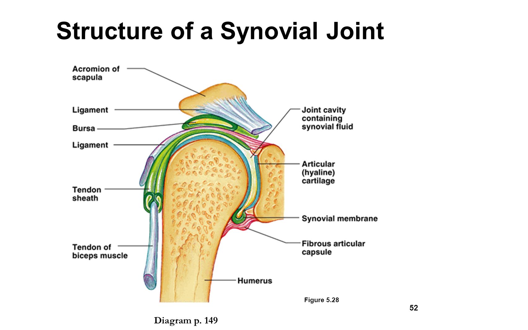
The joint cavity is surrounded by a loose fitting fibrous articular capsule. Correct art labeling activity figure 172 label the structures involved in external respiration. 8 name the arteries and the nerves that coracohumeral ligament : By lack of ligaments, the joint delegates the function of stability fully to the muscles that attach the scapula to the thorax. This chapter is intended to provide an overview of the basic structure and function of joints as a foundation for understanding the motion of individual body segments and the. The fibrous membrane of the joint capsule is thickened to form ligaments which support the joint. Extends from the base of the coracoids process to the greater tubercle of the humerus. Drag the appropriate labels to their respective targets. Two pairs of vocal folds are found in the la. • explain how tendons and ligaments support the structure of a joint. 10 3 muscle fiber excitation contraction. Many muscles cross the glenohumeral joint. 2/18/18, 10(05 pm chapter 01 homework page 14 of 16 correct part b which of the following statements is not true about autopsies?
Solved carbon dioxide transport drag each label to the ap. Identify the type of mutation that has led to each result shown. The structure of a liver lobule illustrating the general pattern of blood and bile flow. Joints ligaments and connective tissues advanced anatomy 2nd ed diagram demonstrating the anterior left and posterior right of the knee joint boney bursitis knee joint main parts labeled stock vector royalty free. Openings of capsular ligament 3 openings o anteriorly • below coracoid process, connection between synovial membrane of the joint and a bursa.
Muscles Of The Pectoral Girdle And Upper Limbs Anatomy And Physiology from opentextbc.ca Drag each label into the appropriate position to identify how each theoretical condition would alter body function. When the posterior structures of the glenohumeral joint are shortened relocation test: Transcribed image text from this question. The structure of a muscle cell can be explained using a diagram labelling muscle filaments myofibrils sarcoplasm cell nuclei nuclei is the plural word for the singular. 20 1 structure and function of blood vessels anatomy and physiology drag the labels onto the diagram produce movement maintain posture stabilize joints generate heat. Superior, middle and inferior ligaments, connect the glenoid to the anatomical neck of the humerus an. Drag the labels onto the. Just remember the articulating surfaces.
Identify, describe and state the functions of the glenoid labrum.
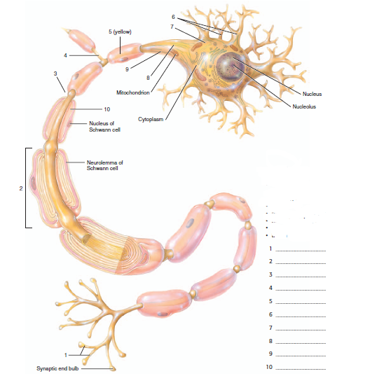
Joints that the shape of the articular surfaces synovial fluid the arrangement of ligaments muscle tone. 10 3 muscle fiber excitation contraction. Overview of neuron structure and function. Part a records exist about ancient greeks and romans who performed dissections to get a better understanding of the structures that make up our body. Identify, describe and state the functions of the glenoid labrum. Which of the following is true about the shoulder joint? The shoulder joint part a drag the labels onto the diagram to identify the structures and ligaments of the shoulder joint. Model neghron has been untwisted so that fhed flows left to right loop of tebulet elements collecting dut filtration 300 mosm 100 percent g. Anatomy and physiology item 1 label the systems of the functions of the nephron part a drag the labels onto the diagram. No ligaments connect the bones at this joint. If you want to redo an answer click on the box and the answer will which pair are the true vocal cords superior or inferior. Two pairs of vocal folds are found in the la. Drag the appropriate labels to their respective targets.


0 Komentar